Once food passes though this region, it enters the cardiac region. Irritation in this area due to fine particle size, stress or other environmental factors can contribute to ulcer formation in swine. This region of the stomach does not secrete digestive enzymes but has significance in that this is where ulcer formation in pigs occurs. The oesophageal region is located at the entrance of the stomach from the oesophagus. The stomach has four distinct areas which include the oesophageal, cardiac, fundic and pyloric regions (Figure 2). The stomach is a muscular organ responsible for storage, initiating the breakdown of nutrients, and passing the digesta into the small intestine. Movement though the oesophagus involves muscle peristalsis, whichis the contraction and relaxation of muscles to move the food. Once food is chewed and mixed with saliva, it passes though the mouth, pharynx and then the oesophagus to the stomach. The contribution of digestive enzymes from saliva is minor but still noteworthy. Saliva generally contains very low levels of amylase, the enzyme that hydrolyses starch to maltose. Thereby in a dry diet, more saliva mucus is secreted while in a moist diet, only an amount to assist with swallowing is secreted. The amount of mucus present in saliva is regulated by the dryness or moistness of the food consumed. Saliva secretion is a reflex act stimulated by the presence of food in the mouth. There are three main salivary glands, which include the parotid, mandibular and sub-lingual glands. While teeth serve the main role in grinding to reduce food size and increase surface area, the first action to begin the chemical breakdown of food occurs when feed is mixed with saliva. The mouth serves a valuable role not only for the consumption of food but it also provides for the initial partial size reduction though grinding.

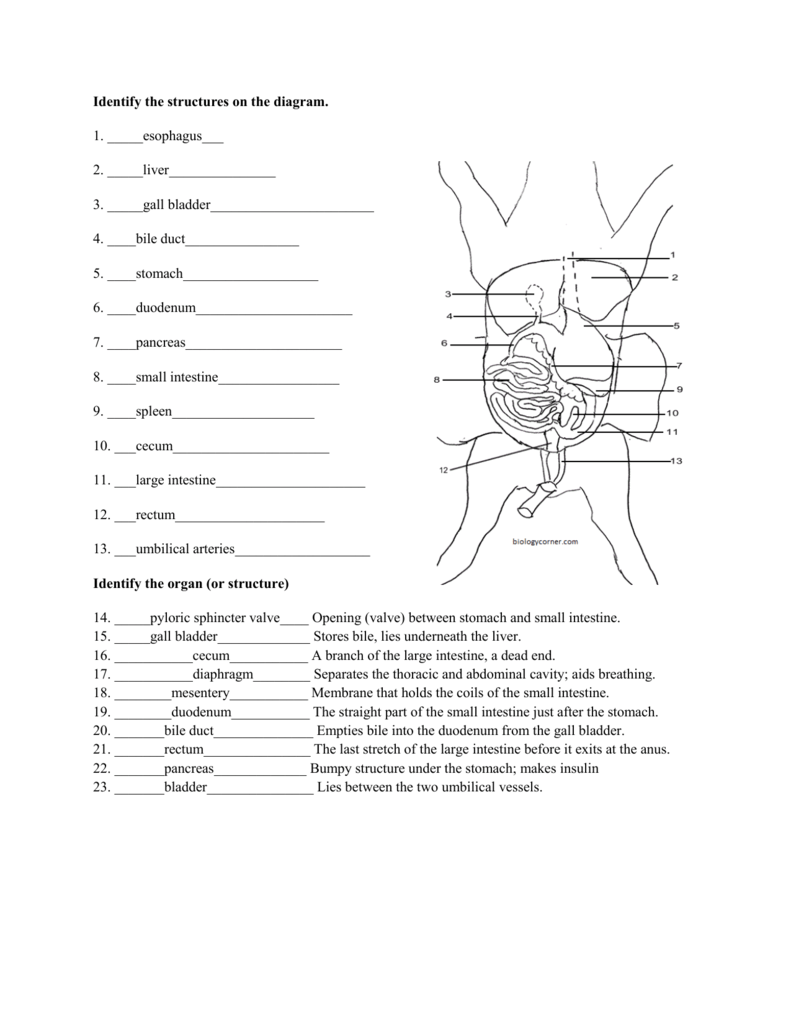
I think what they want is duodenum 7.Īs the pig is a mammal many aspects of its structural and functional.Figure 1.

Start studying fetal pig dissection hand in. Use the following fetal pig diagrams to review the anatomy of a fetal pig. The first one above is the photograph of a real dissected pig. While this page summarizes the information needed for the lab practicum a very good site for further review can be found at the following.
Pig dissection diagrams manual#
Before observing internal or external structures of the fetal pig use your dissection manual textbook and dissection notebook to answer the pre lab questions on the fetal pig.
Pig dissection diagrams free#
Mark stanback You are free to modify these documents. Fetal pig dissection with photos developed by dr. Appendix or ceccum i really cant tell from the diagram 11. Learn vocabulary terms and more with flashcards games and other study tools.

Fetal pig dissection lab answers introduction pigs one of the most similar animals to humans have been used to inform and teach students about the circulatory respiratory and digestive system through a procedure called a dissection for many years. The student handouts are available in these formats.

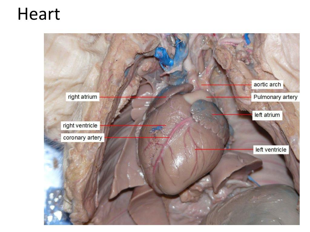
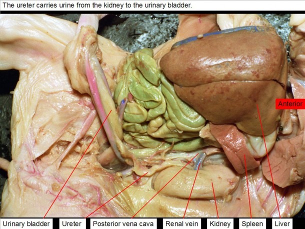
These pictures of fetal pig dissection are useful for learning the anatomy of the fetal pig.įetal Pig Dissection Bingle Vet No idea what its point to 5.įetal pig dissection diagram answers. Preserved fetal pig dissecting pan dissecting kit dissecting pins string plastic bag metric ruler paper towels.


 0 kommentar(er)
